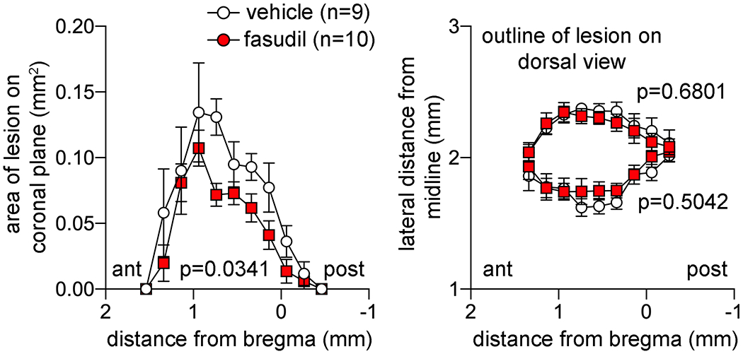
Figure 4: Lesion morphometry.

Lesion areas on coronal plane (left) and distance from the midline of the medial and lateral lesion boundaries at 1 month are plotted along the anteroposterior axis. Two-way ANOVA for repeated measures.

Lesion areas on coronal plane (left) and distance from the midline of the medial and lateral lesion boundaries at 1 month are plotted along the anteroposterior axis. Two-way ANOVA for repeated measures.