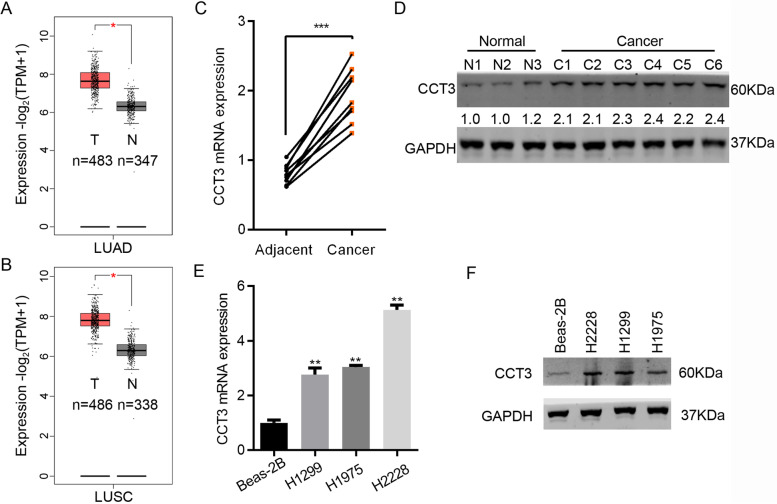
Fig. 1.
CCT3 is overexpressed in lung cancer tissues and cells. A The transcript abundance of CCT3 was analyzed in lung adenocarcinoma (LUAD, n = 483) and normal tissues (n = 347) from TCGA database. *p < 0.05. B The transcript abundance of CCT3 was analyzed in lung squamous cell carcinoma (LUSC, n = 486) and normal tissues (n = 338) from TCGA database. *p < 0.05. C Nine pairs of lung cancer and adjacent normal tissues were subjected to qRT-PCR analysis of CCT3 expression. ***p < 0.001. D Western blot analysis of CCT3 in lung cancer and paired adjacent normal tissues. The blots were cut prior to hybridisation with antibodies during blotting. E and F qRT-PCR (E) and Western blot (F) analysis of CCT3 in normal lung cells Beas-2B and in cancer cells H2228, H1299 and H1975. **p < 0.01. GAPDH is the internal control. The blots were cut prior to hybridisation with antibodies during blotting

