Fig. 5.
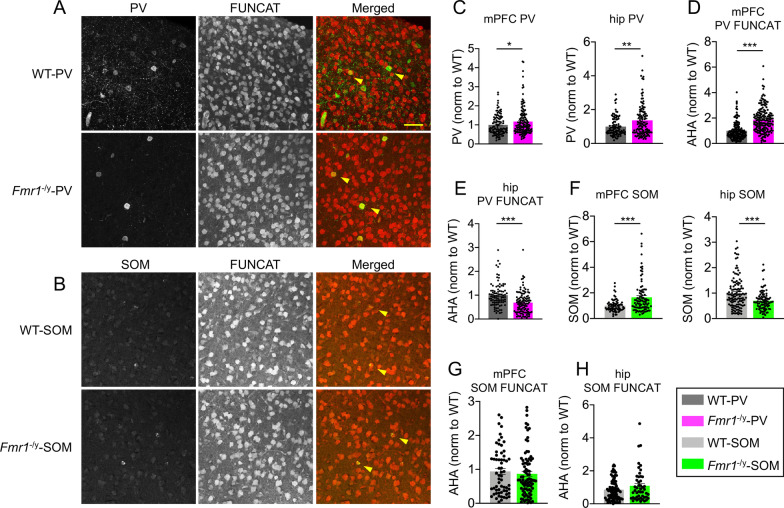
Brain region-specific cellular deficits in PV- and SOM-positive neurons upon cell type-specific deletion of Fmr1. A, B Representative images of FUNCAT and PV staining from WT-PV and Fmr1−/y-PV mouse mPFC (A, scale bar: 50 μm) or FUNCAT and SOM staining from WT-SOM and Fmr1−/y-SOM mice (B). Arrows denote individual PV- or SOM-expressing cells with FUNCAT signal. C PV expression was elevated in mPFC and hippocampus of Fmr1−/y-PV mouse. Quantification of PV labeling in PV-positive neurons in WT-PV and Fmr1−/y-PV mouse mPFC and hippocampus. D, E Cell type-specific deletion of Fmr1 results in elevated de novo protein synthesis in PV-positive neurons in mPFC, but a decrease in hippocampus of Fmr1−/y-PV mice. Quantification of AHA fluorescence in PV-positive neurons in WT-PV and Fmr1−/y-PV mouse mPFC D and hippocampus E. Values represent mean ± SEM (hippocampus, n = 45–48 z-stacks, 19–24 sections, from 3 animals per genotype; mPFC, n = 24–25 z-stacks, 14 sections, from 3 animals per genotype); ***p < 0.0001 (Student’s t test). F SOM expression was elevated in mPFC but decreased in hippocampus of Fmr1−/y-SOM mice. Quantification of SOM labeling in SOM-positive neurons in WT-SOM and Fmr1−/y-SOM mouse mPFC and hippocampus. G, H De novo protein synthesis is not significantly different upon cell type-specific deletion of Fmr1 in SOM-positive neurons. Quantification of AHA fluorescence in SOM-positive neurons in WT-SOM and Fmr1−./y-SOM mouse mPFC G and hippocampus (H). Values represent mean ± SEM (hippocampus, n = 26–34 z-stacks, 11–14 sections, from 2 animals per genotype; mPFC, n = 39 z-stacks, 21–22 sections, from 3 animals per genotype); ***p < 0.0001 (Student’s t test)

