Figure 1.
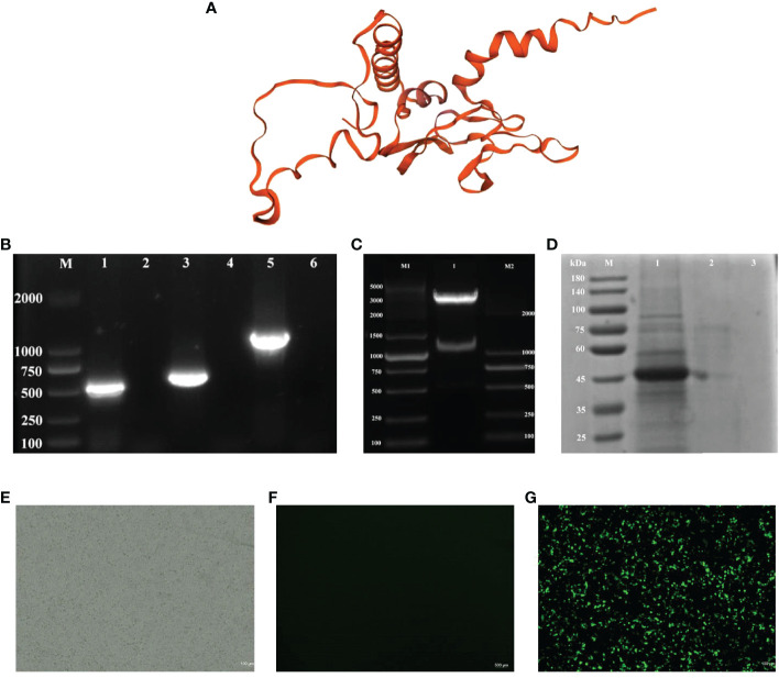
Identification of pPG-612-CK6-G/L.casei 393 and the protein expression. (A) Three-dimensional structures of recombinant CK6 and G proteins (B) Electrophoresis plots of amplification of different target genes, line 1: the G gene strip; line 3: the CK6 gene strip; line 5: the recombinant CK6-G strip; line 2, 4 and 6: blank control; (C) Double digestion electrophoresis plots of recombinant plasmids, 1: recombinant plasmid double digestion strip; M1: DNA marker DL5000, M2: DNA marker DL2000; (D) SDS-PAGE identification of protein expression of CK6-G gene, line 1: the pPG-612-CK6-G/L.casei 393 after induction; line 2: the uninduced pPG-612-CK6-G/L.casei 393; 3: blank control; M: 10-190 kDa protein marker (E) Indirect immunofluorescence map of the blank group. (F) Indirect immunofluorescence map of the control group. (G) Indirect immunofluorescence plot of the experimental group.

