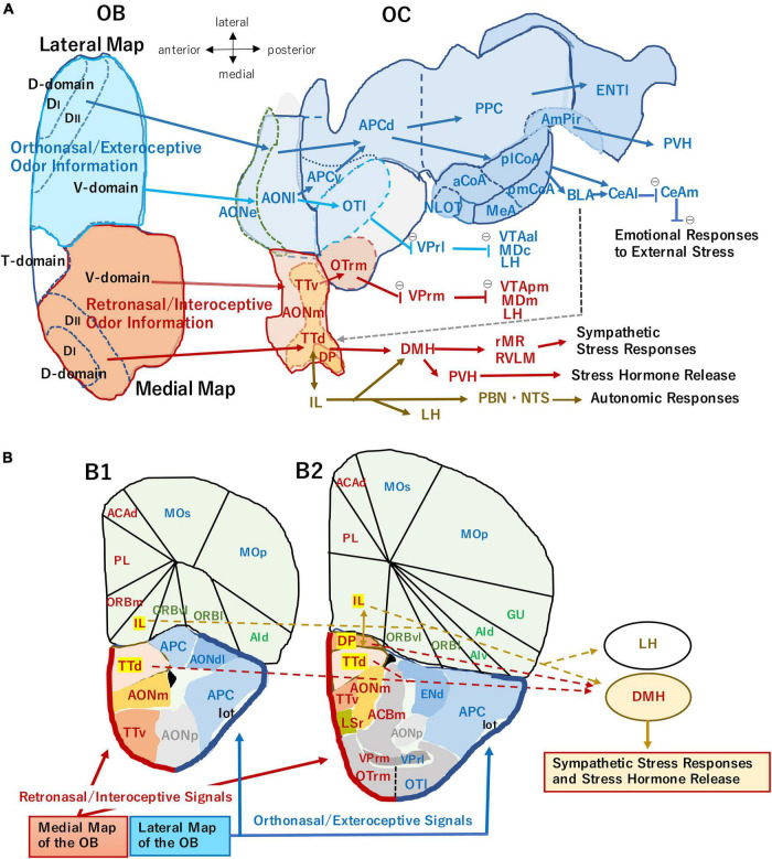
FIGURE 1.
(A) An unfolded map (ventral center view) of the mouse olfactory bulb (OB) and olfactory cortex (OC) illustrating two olfactory processing streams. The medial stream (magenta arrows) originates from the medial map of the OB (dark orange) and conveys retronasal/interoceptive information to the medial part of the anterior olfactory nucleus (AONm), ventral tenia tecta (TTv), dorsal tenia tecta (TTd), dorsal peduncular cortex (DP), and rostromedial part of olfactory tubercle (OTrm). OTrm sends inhibitory axons (marked by θ) to the rostromedial part of ventral pallidum (VPrm). The VPrm projects inhibitory axons to the posteromedial part of ventral tegmental area (VTApm), medial part of mediodorsal nucleus in the thalamus (MDm), and lateral hypothalamus (LH). The TTd and DP project to the dorsomedial nucleus of hypothalamus (DMH), which projects to the sympathetic premotor neurons in the rostral medullary raphe region (rMR) and rostral ventrolateral medulla (RVLM) to generate sympathetic responses. The TTd and DP have reciprocal connections with infralimbic cortex (IL), which projects to the autonomic centers in the hypothalamus and brainstem, such as DMH, LH, parabrachial nucleus (PBN), and nucleus of solitary tract (NTS). The ventral part of DMH projects to the paraventricular nucleus of hypothalamus (PVH) and activates the hypothalamo-pituitary-adrenal axis to release stress hormones (Herman et al., 2016; Nakamura and Morrison, 2022). The lateral stream (blue arrows) originates from the lateral map of the OB (blue) and sends orthonasal/exteroceptive odor information to the external and lateral parts of the anterior olfactory nucleus (AONe, AONl), lateral part of the olfactory tubercle (OTl), ventral and dorsal parts of the anterior piriform cortex (APCv, APCd), posterior piriform cortex (PPC), nucleus of the lateral olfactory tract (NLOT), anterior, posterolateral, and posteromedial nuclei of the cortical amygdala (aCoA, plCoA, pmCoA), medial amygdala (MeA), amygdalo-piriform transition area (AmPir), and lateral entorhinal area (ENTl). The CoA projects to the basolateral amygdaloid complex (BLA) and lateral and medial nuclei of central amygdala (CeAl, CeAm) to induce emotional responses. A gray broken arrow from the amygdala to the DP/TTd indicates a putative route for the transmission of aversive orthonasal odor information to the DP/TTd-DMH pathway. The OTrl sends inhibitory axons to the rostrolateral part of ventral pallidum (VPrl). The VPrl projects inhibitory axons to the anterolateral part of ventral tegmental area (VTAal), the central part of mediodorsal nucleus in the thalamus (MDc), and LH. The unfolded map was modified from Mori and Manabe (2014), with permission from Springer Nature. (B) Medial and lateral areas of the OC shown on the coronal section through the olfactory peduncle (B1) and that through the rostral part of the OT (B2). Thick magenta line indicates the superficial layer of the medial OC mainly receiving retronasal information from the medial OB map. Thick blue line indicates the superficial layer of the lateral OC mainly receiving orthonasal information from the lateral OB map. The IL, DP, and TTd (highlighted) form the ventromedial prefrontal cortex, and their projection to the hypothalamus is shown by broken arrows. ACAd, dorsal part of anterior cingulate area; ACBm, medial part of accumbens nucleus; AId, dorsal part of agranular insular cortex; AIv, ventral part of agranular insular cortex; AONdl, dorsolateral part of anterior olfactory nucleus; AONp, posterior part of anterior olfactory nucleus; ENd, dorsal endopiriform nucleus; Gu, gustatory cortex; lot, lateral olfactory tract; LSr, rostral part of lateral septum; MOp, primary motor cortex; MOs, secondary motor cortex; ORBl, lateral part of orbital cortex; ORBm, medial part of orbital cortex; ORBvl, ventrolateral part of orbital cortex; OTl, lateral part of olfactory tubercle; OTrm, rostromedial part of olfactory tubercle; PL, prelimbic area; VPrl, rostrolateral part of ventral pallidum; VPrm, rostromedal part of ventral pallidum. The contour of each area is generated using Allen Brain Atlas (https://atlas.brain-map.org), the Allen Institute for Brain Science (2004).

