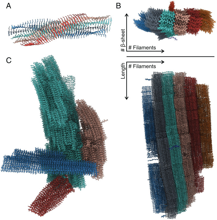
Fig. 3.
(A) A filament model observed at the end of a simulation. Colors indicate the five different β-sheets composing the filament. (B) Top and side view of a mature fibril with colors indicating the different filaments. From the top view, it is possible to see a peptide which is about to attach to the fibril and a protofilament which is perpendicular to the main fibril. (C) Multiple mature fibrils (each represented with a different color) interacting with each other from one of the 13-mM simulations.

