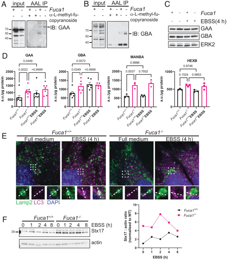
Fig. 4.
FUCA1 regulates fucosylation status and activities of lysosomal enzymes, and promotes autophagosome–lyosome fusion. (A–D) Primary Fuca1flox/flox MEFs were infected with a retrovirus expressing the Cre recombinase (Fuca1−/−) or a retrovirus control empty vector (Fuca1+/+). Cell lysates were immunoprecipitated using biotinylated AAL, preincubated with methyl-α-l-fucopyranoside as a control (A and B). Eluted proteins were then loaded on a gel for immunoblotting analysis using antibodies directed against GAA (A) and GBA (B). Whole-cell lysates (input) were analyzed using the same antibodies. (C) Primary Fuca1+/+ and Fuca1−/− MEFs were incubated in EBSS for 4 h. Cell lysates were analyzed by Western blot using antibodies against GAA, GBA, and ERK2, which was used as a loading control. (D) GAA, GBA, MANBA, and HEXB enzymatic activities were assessed in Fuca1+/+ and Fuca1−/− MEFs under baseline and starvation conditions, and expressed as arbitrary units (a.u.) per microgram of total protein (n = 3 independent experiments, one-way ANOVA with Bonferroni multiple comparison test). (E) Subcellular localization of LC3B and Lamp2 was visualized in immortalized Fuca1+/+ and Fuca1−/− MEFs under baseline conditions and after 4-h EBSS starvation by confocal microscopy. Nuclei were stained with DAPI. (Scale bars, 5 µm.) Regions of interest are highlighted in white boxes. (F) Immortalized Fuca1+/+ and Fuca1−/− MEFs were incubated in EBSS for the indicated time period. Cell lysates were analyzed by Western blot using Stx17 and actin antibodies. The Stx17 : actin ratio is plotted on the right.

