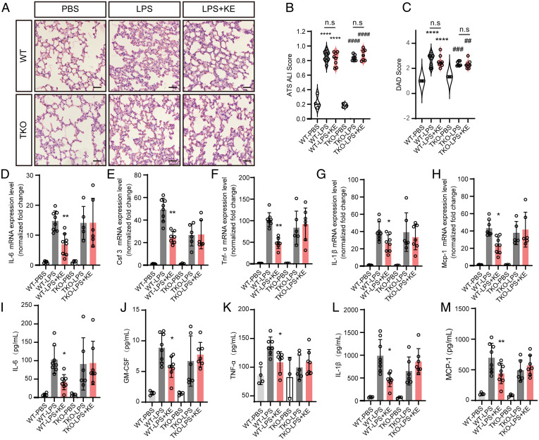
Fig. 7.
Agonistic activation of Tas2R by KE-A attenuates LPS-induced acute pulmonary inflammation. (A) The histology of the lung tissues of LPS- or PBS-inhaled mice with and without KE-A treatment (H&E staining, 20×). (Scale bar, 50 μm.) The alveolar walls of PBS-inhaled mice are very thin, and the alveoli contain no infiltrated cells, while the LPS-inhaled lungs show apparent inflammatory infiltration. Transient treatment with KE-A does not change such inflammation infiltration and other pulmonary structures. Blinded histopathological evaluation of lung damage was performed by using the acute lung injury (ALI) scoring systems (B) and defuse alveolar damage (DAD) histological scoring systems (C) that were generated by the American Thoracic Society. ****P < 0.0001, * compared with WT PBS ; ##P < 0.01, ###P < 0.001, ####P < 0.0001, # compared with TKO PBS; n.s.: no significance. Two-way ANOVA. (D–H) The expression levels of inflammation-associated genes of mice (n = 3 to 8). (I–M) The concentrations of the multiple inflammation cytokines in the lung lysates were measured by a Luminex assays kit (n = 3 to 8). *P < 0.05, **P < 0.01, compared with WT LPS group; two-way ANOVA.

