
Figure 15:
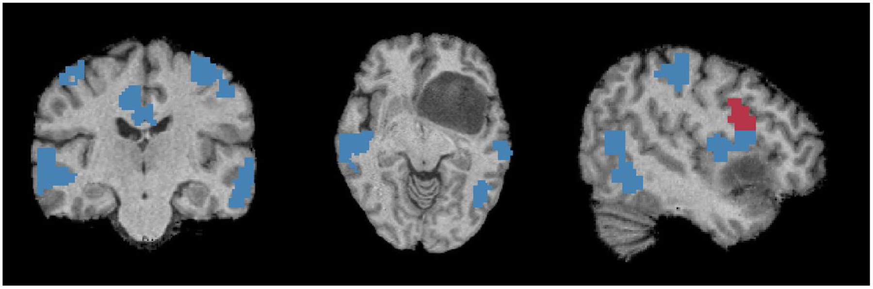
From (L-R) we show coronal, axial and saggital views of correct (blue) and incorrect (red) prediction by our model for the eloquent cortex in a challenging inferior frontal gyrus tumor case.
Official websites use .gov
A
.gov website belongs to an official
government organization in the United States.
Secure .gov websites use HTTPS
A lock (
) or https:// means you've safely
connected to the .gov website. Share sensitive
information only on official, secure websites.
From (L-R) we show coronal, axial and saggital views of correct (blue) and incorrect (red) prediction by our model for the eloquent cortex in a challenging inferior frontal gyrus tumor case.