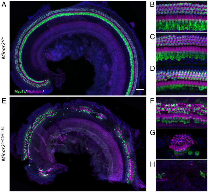
Fig. 6.
Hair cell degeneration pattern in Minar2tm1b mutant mice at 14 wk old. Confocal maximum intensity projection images of the whole-mount organ of Corti of 14-wk-old mice. Hair cells were examined using anti-Myo7a antibody (green) and phalloidin (magenta). Nuclei are shown in blue (DAPI). (Scale bars: 200 µm in A and 10 µm in B.) The images are representative examples of Minar2+/+ (A–D) and Minar2tm1b/tm1b (E–H) mice. (A and E) Overview of the apical half of the cochlea. (B–D) High-magnification images corresponding to the 12 kHz, 24 kHz, and 36 kHz best-frequency regions, respectively. (F–H) High-magnification images of three different patterns of hair cell loss observed in Minar2tm1b mutant mice. All five homozygotes that were studied at 14 wk old showed similar patches of loss of the organ of Corti at varying locations along the length of the cochlear duct interspersed with regions that showed only scattered hair cell loss. Minar2+/+ (n = 4), Minar2+/tm1b (n = 5), and Minar2tm1b/tm1b (n = 5).

