Figure 3.
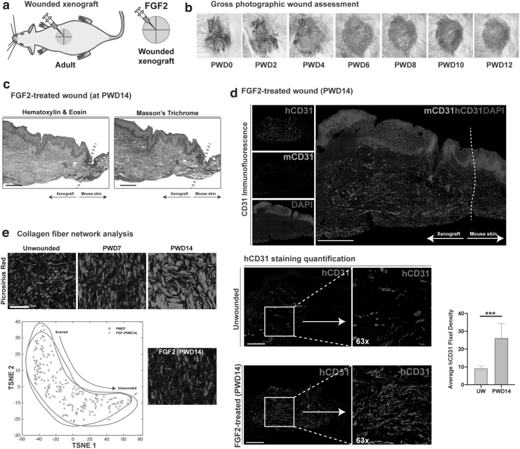
The xenograft model is a platform for exploring the efficacy of topical agents. (a) Graphical representation of the wounded and unwounded xenograft with FGF treatment. (b) Representative gross images of a FGF2-treated wound at PWDs 0 through 12. (c) Representative FGF2-treated wounded xenograft at PWD14 stained with H&E and Masson's Trichrome. Scale bar: 200 μm. (d) Immunofluorescence staining for mouse- (green) and human-specific (red) endothelial cells (CD31/PECAM). Scale bar: 200 μm. Quantitative comparison of mean pixels positive for hCD31 in unwounded and FGF2-treated wounds at PWD14. ***p < 0.001. (e) 2D TSNE plots of collagen fiber networks in PWD7 (gray) and FGF2-treated wounds at PWD14 (black outline), showing overlap with no distinct difference. FGF2, fibroblast growth factor 2.

