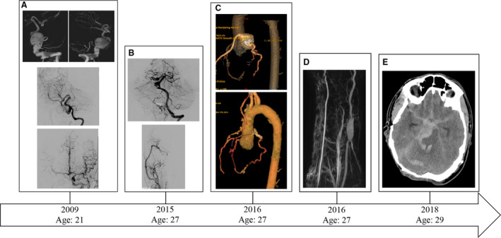
Figure 1. Radiographic correlate of the index patient’s clinical course.

A, (Top row) Digital subtraction angiography showing 3D reconstruction of a right vertebral artery fusiform aneurysm after a right vertebral injection. A fusiform aneurysm encompassing the extracranial and intracranial vertebral artery is seen. (Middle) Post‐treatment left vertebral artery injection shows a normal left vertebral artery and small fusiform dilatation of the right P1 segment. The right vertebral artery underwent coil embolization/sacrifice with bypass. (Bottom) Digital subtraction angiogram with Townes view of a left common carotid artery injection. The right anterior circulation is seen due to cross‐filling via a patent anterior communicating artery. No aneurysmal pathology is identified in the anterior circulation. The right common carotid artery had previously undergone coil embolization and sacrifice. B, Follow‐up digital subtraction angiography showing (Top) progression of the right P1 segment fusiform aneurysm as well as development of a mid‐basilar artery aneurysm and (Bottom) right external carotid artery to middle cerebral artery bypass with normal right middle cerebral artery candelabra. C, (Top) Identification of a coronary artery aneurysm and (Bottom) post‐treatment imaging. D, Identification of a right radial artery fusiform aneurysm. E, Non‐contrasted computed tomography showing diffuse subarachnoid hemorrhage secondary to a ruptured basilar artery aneurysm causing death.
