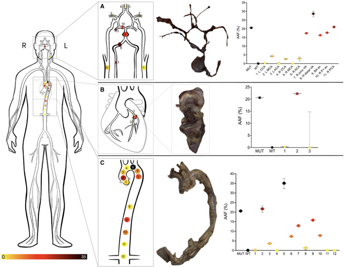
Figure 2. Heatmap of vascular samples harvested from the (A) intracranial, (B) coronary, and (C) aortic samples.

Gross pathological specimens are shown as well as AAF% at the associated sampling sites.

Gross pathological specimens are shown as well as AAF% at the associated sampling sites.