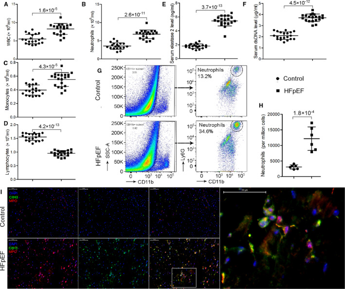
Figure 1. Neutrophils and NETs markers are elevated in patients and mice with HFpEF.

A through D, Patients with HFpEF showed significantly higher levels of circulating white blood cells (A), particularly neutrophils (B) and monocytes (C) but not lymphocytes (D) compared with matched healthy controls. E through F, Circulating NETs markers, that is, elastase 2 (E) and cell‐free dsDNA (F) were significantly higher in the sera of patients with HFpEF compared with healthy controls. G through H, Flow cytometric analysis showed that HFpEF mice had an over 3‐fold increase in neutrophil counts in the heart. I, In situ immunofluorescence identifying NETs by extracellular MPO (red), CitH3 (green), and DNA (blue) deposits in the heart, showing increased NETs formation in HFpEF mice than controls. Scale bar represents 50 µm. CitH3 indicates citrullinated histone 3; dsDNA, double‐stranded DNA; HFpEF, heart failure with preserved ejection fraction; MPO, myeloperoxidase; NETs, neutrophil extracellular traps; and WBC, white blood cell.
