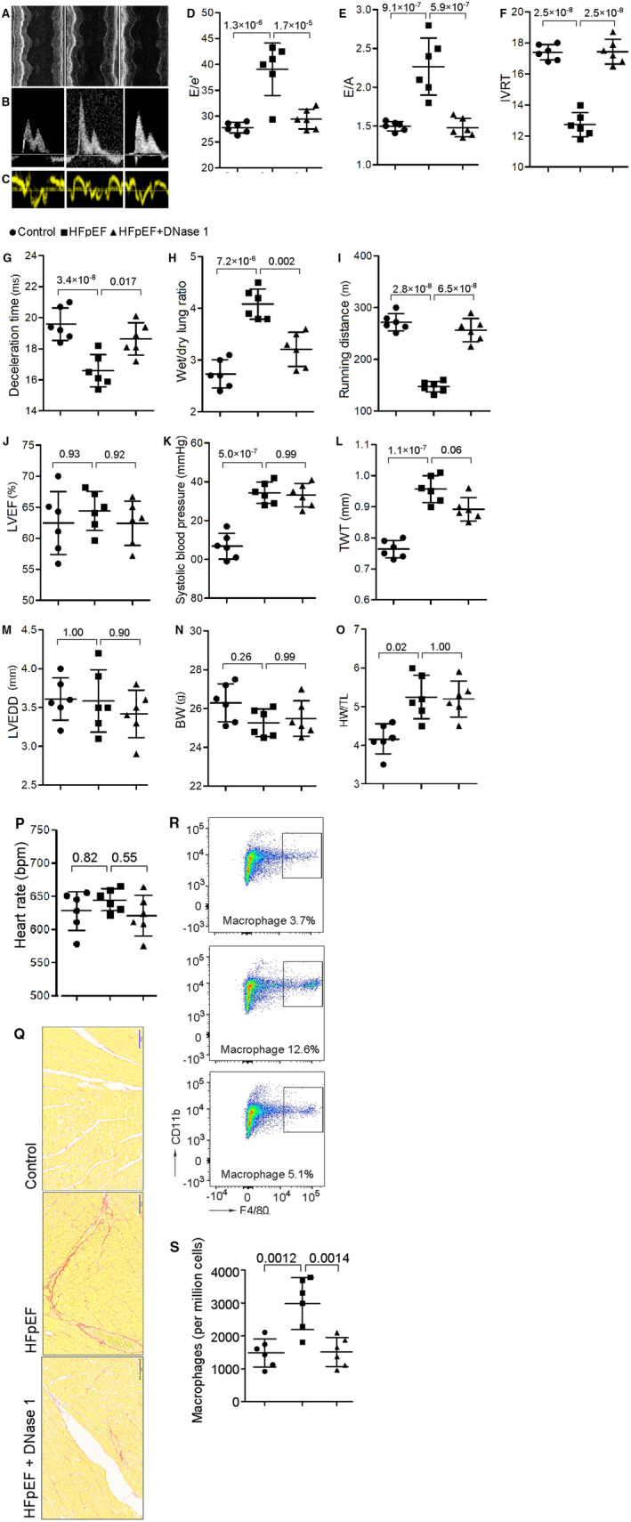
Figure 2. NETs inhibition reduces cardiac macrophage infiltration, attenuates cardiac fibrosis, and ameliorates diastolic function in HFpEF mice.

A through C, Representative echocardiography images of left ventricular M‐mode echocardiography (A), pulsed‐wave Doppler (B) and tissue Doppler (C) tracings. D, Ratio between mitral E wave and e′ wave (E/e′). E, Ratio between mitral E wave and A wave (E/A). F, Isovolumic relaxation time (IVRT). G, Deceleration time; H, Ratio between wet and dry lung weight. I, Running distance during exercise exhaustion test. J, Percentage of LVEF. K, Systolic blood pressure. L, Total wall thickness (TWT). M, Left ventricular end‐diastolic diameter (LVEDD). N, Body weight (BW). O, Ratio between heart weight and tibia length (HW/TL). P, Heart rate. These results showed that DNase 1 significantly attenuated diastolic dysfunction, improved exercise tolerance, reduced lung congestion, but had no significant effect on LVEF, blood pressure or cardiac hypertrophy. Q, Representative images of Sirius red staining of heart tissue, showing an increment of cardiac fibrosis in HFpEF and an attenuation after DNase 1 treatment. Scale bar represents 50 µm. R and S, Flow cytometric analysis showing that HFpEF mice had a significant increase in macrophage counts in the heart, and NETs inhibition with DNase 1 treatment significantly reduced the number of macrophages in the heart. BW indicates body weight; DNase 1, deoxyribonuclease; HFpEF, heart failure with preserved ejection fraction; LVEF, left ventricular ejection fraction; NETs, neutrophil extracellular traps; and TWT, total wall thickness.
