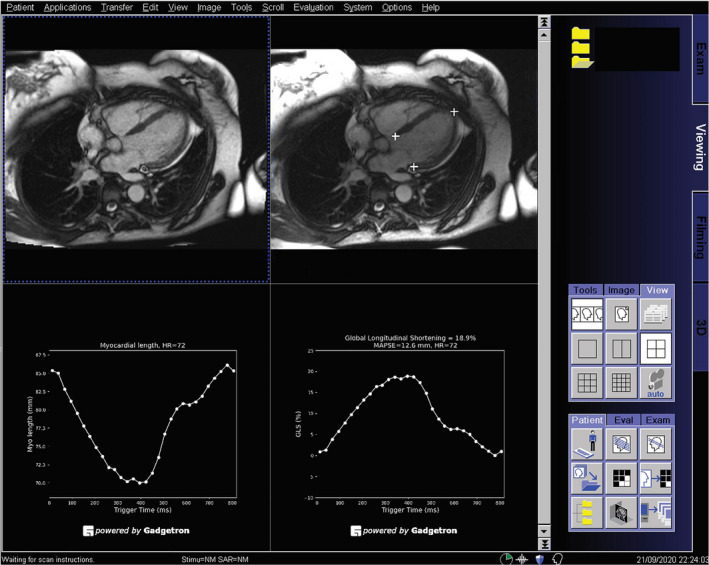
Figure 2. The proposed artificial intelligence solution was deployed to the magnetic resonance (MR) scanners in the in‐line fashion illustrated by this screen snapshot of the MR scanner console, where a 4‐chamber cine series was processed with the model.

The cine images are displayed at the top left, and detected landmarks are overlaid on the images at the top right. This allows a convenient check of performance of landmark detection. The global longitudinal shortening (GL‐Shortening) and mitral annular plane systolic excursion (MAPSE) were measured automatically and displayed as signal curves. In this way, a fully automated solution was achieved, without requiring any user interaction for processing.
