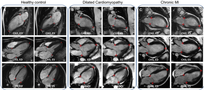
Figure 3. Examples of artificial intelligence detection on long‐axis cine images.

The detected landmarks were overlaid on the image to indicate the accuracy of the model. A, A healthy 31‐year‐old man from the scan–rescan cohort was scanned to acquire 3 long‐axis cine views. B, A 61‐year‐old woman was diagnosed with dilated cardiomyopathy. C, A 30‐year‐old man with myocardial infarction (MI) was scanned and found to have impaired cardiac function with the ejection fraction being 22%. CH2 indicates 2‐chamber; CH3, 3‐chamber; CH4, 4‐chamber; ED, end‐diastolic; and ES, end‐systolic.
