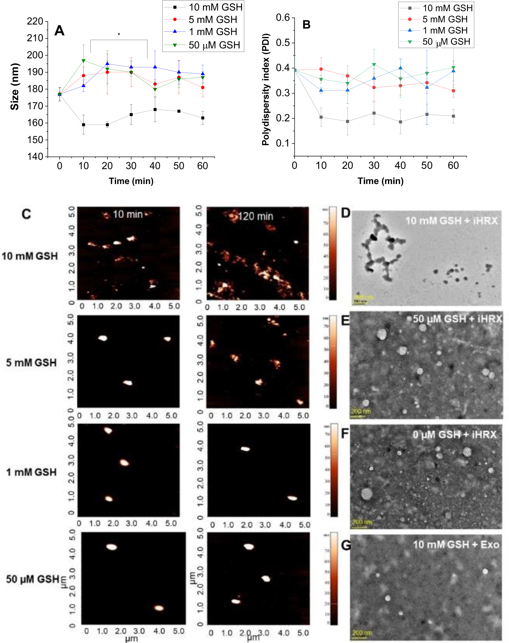
Figure 7.

Size and shape of iHRX in the presence of glutathione. (A) Hydrodynamic diameters of HRX from 0 – 120 min in 10 min increments with increasing amounts of glutathione. (B) polydispersity indices of HRX from 0 – 120 min in 10 min increments with increasing amounts of glutathione. (C) AFM images of HRX at 10 min (left) and 120 min (right). (D) HR-TEM images of iHRX with 10 mM glutathione at 120 min. (E) HR-TEM images of iHRX with 50 μM glutathione at 120 min. (F) HR-TEM images of iHRX with 0M glutathione at 120 min. (G) HR-TEM images of exosomes with 10 mM glutathione at 120 min.
