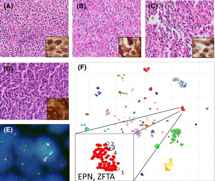
FIGURE 4.

Undifferentiated brain tumors with ZFTA fusion. Histomorphology of four examples (cases 1–4) is shown in A–D, respectively, with insets demonstrating nuclear expression of p65 as a marker for the activation of NFκB signaling. Break‐apart of RELA (arrow in E) as demonstrated by FISH analysis (exemplarily shown for case 1) as well as t‐SNE analysis after methylation profiling (F) further suggest the diagnosis of a supratentorial ependymoma with ZFTA fusion. F shows reference data from 2800 brain tumors previously published by Capper et al. as well as the four cases shown in A–D
