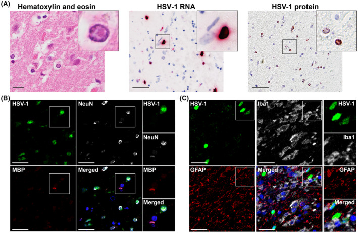
FIGURE 2.

Lytic HSV infection of neurons in the brain of herpes simplex virus encephalitis (HSE) patients. (A) Brain sections from HSE patients (Table 1) were stained with hematoxylin and eosin, or stained for HSV‐1 LAT RNA by ISH and HSV‐1 ICP8 protein by IHC. Boxes indicate the area shown at higher magnification in the inset. Scale bar: 50 µm. (B, C) Brain sections of HSE patients were immunofluorescently stained for (B) HSV‐1 protein (green), neurons (NeuN; white) and oligodendrocytes (MBP; red), as well as (C) HSV‐1 protein (green), microglia (Iba1; white), and astrocytes (GFAP; red). Nuclei were stained with Hoechst‐33342 (blue). Representative images are shown for patient #2 (amygdala/entorhinal cortex; Table 1). Scale bar: 20 µm
