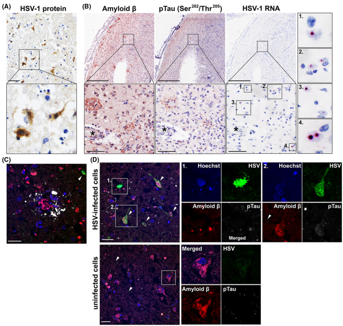
FIGURE 5.

Detection of HSV‐infected cells in close proximity to Aβ plaques in the brain of an Alzheimer's disease patient with herpes simplex encephalitis. (A) Brain section stained for HSV protein (brown) by immunohistochemistry (IHC). Scale bar indicates 50 µm. (B) Consecutive brain sections were stained for Aβ (4G8) and pTau (Ser202, Thr205) by IHC or stained for HSV‐1 RNA by in situ hybridization (ISH). Boxes indicate areas shown at higher magnification. Asterisks indicate the same blood vessel in consecutive sections. Scale bars indicate 500 µm (low magnification) and 100 µm (high magnification). (C, D) Brain sections were immunofluorescently stained for HSV protein (green), Aβ (red), and pTau (Ser202/Thr205; white) protein. Nuclei were stained with Hoechst‐33342 (blue). Scale bar: 20 µm
