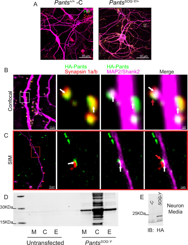
Fig 2. Synaptic localization and release of Pants.
A) Immunocytochemistry with HA antibody shows Pants localization throughout cultured hippocampal neurons from PantsSOG-Y/+ mice, but not wildtype controls. B) Immunocytochemistry of 18–21 div primary neurons from PantsSOG-Y hippocampus stained for HA, a Pre-synaptic marker (Synapsin 1a/b) and post-synaptic markers (MAP2 and Shank2) imaged with confocal microscopy. White arrows indicate areas of colocalization, and red arrows indicate suspected extra-synaptic pants that fails to co-localize with any markers. C) Structured Illumination Microscope imaging of the same staining from (A) to obtain better resolution. D) HA Western blot of HA IP from Nero2a cellular lysates (C), extracellular matrix (E) fractions, and proteins precipitated from cell media (M) after transfection with a construct for over-expressing the PantsSOG-Y fusion protein compared to untransfected controls. E) HA Western blot of HA IP of media collected from primary PantsSOG-Y hippocampal neurons or Pants+/+ negative control neurons.

