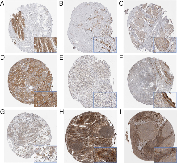
Fig 2. Immunohistochemistry staining for CCBL2.
The expression of CCBL2 in breast cancer (B and C), renal cancer (E), ovarian cancer (G) and head and neck cancer (I) cells were decreased than in respective normal tissues (A, D, F and H). In breast cancer, infiltrating lobular carcinoma (B) expressed more CCBL2 than infiltrating ductal carcinoma (C).

