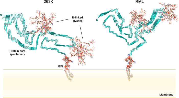
Fig 2. Cross-sectional depictions of membrane-bound 263K and RML prions.
For simplicity, only short pentameric segments of the fibrils are shown. The natural glycans and GPI moieties are much more heterogeneous than those shown. The RML structure shown was assembled using the aRML pdb coordinates (PDB: 7TD6) because the wtRML pdb coordinates were not yet publically available. Although the aRML and wtRML cores are similar overall, there are detailed features of the wtRML core structure that differ from that depicted. See [10]. GPI, glycophosphatidylinositol.

