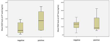
Figure 3.

Boxplots representing significant distributions of median serum VEGF and PDGF levels of patients who had progression on treated liver lobe in the 6th week of follow-up
VEGF: Vascular endothelial growth factor, PDGF: Platelet-derived growth factor, Negative: No progression, Positive: Progression
