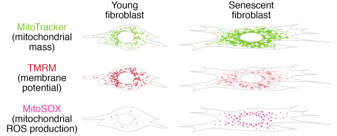
Figure 1. Mitochondrial dysfunction in senescence.
Illustrations are representative of mitochondrial mass, membrane potential, and ROS levels in young versus senescent fibroblasts as observed after staining with fluorescent dyes. Mitochondria in human fibroblasts can be stained with MitoTracker (green) to show mitochondrial mass and tetramethylrhodamine methyl ester (TMRM) (red), which accumulates in mitochondria in a membrane potential–dependent fashion at under-saturated concentrations. There is higher mitochondrial mass in senescent fibroblasts, but their membrane potential is lower (as indicated by weak and patchy TMRM staining) than in non-senescent (young) fibroblasts. The mitochondrial network is more fragmented in young cells, while mitochondria are fused in senescence. Mitochondrial superoxide levels can be visualized using MitoSOX (pink). Mitochondrial superoxide levels are elevated in senescent human fibroblasts.

