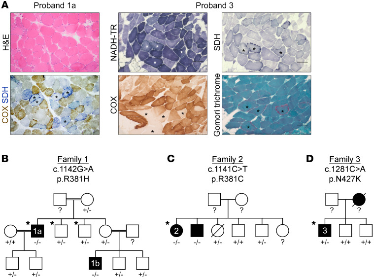
Figure 1. Identification of MDDS and candidate RRM1 variants.
(A) Histology of muscle biopsy cross sections. Asterisks indicate ragged-blue (NADH-TR and SDH), COX-negative, and ragged-red (modified Gomori trichrome) fibers. Scale bar: 100 μm. (B) Family 1, (C) family 2, and (D) family 3 pedigrees, indicating affected individuals (black), WES (asterisks), and Sanger sequencing (+, normal; –, variant; ?, not done).

