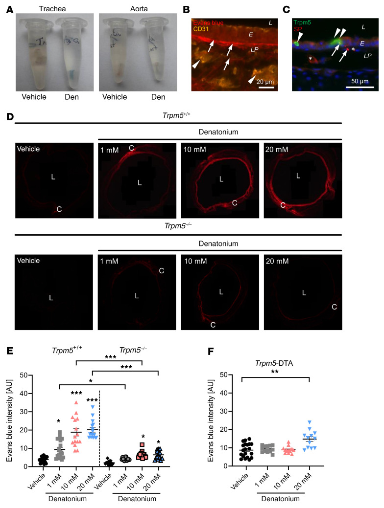
Figure 2. Evans blue (EB) extravasation in response to denatonium.
(A) Explanted tracheae (left) and aortae (right) from mice treated with vehicle control (PBS) or denatonium (den) show blue color due to EB extravasation in the trachea after denatonium treatment. EB is absent in the aorta. (B) CD31 staining with EB fluorescence in murine tracheal sections after denatonium treatment. (C) Costaining against Trpm5 (BCs, arrowheads) and SP (sensory nerve endings) showing blood vessels (*) and nuclei (blue, DAPI). Scale bars: 20 μm (B) and 50 μm (C). (D) Images of tracheal rings showing EB fluorescence of animals treated with PBS (vehicle), 1, 10, or 20 mM denatonium in WT (Trpm5+/+) or Trpm5-knockout (Trpm5–/–) mice. (E) Quantification of EB extravasation in response to 1, 10, or 20 mM denatonium. (F) Quantification of EB extravasation in BC-depleted mice (Trpm5-DTA) in response to denatonium. In E and F, data are shown as single values and mean ± SEM (n = 12–20 rings from 3–4 mice). L, lumen; E, epithelium; LP, lamina propria; C, cartilage. *P < 0.05; **P < 0.01; ***P < 0.001 by 1-way ANOVA followed by Bonferroni’s multiple-comparison correction.

