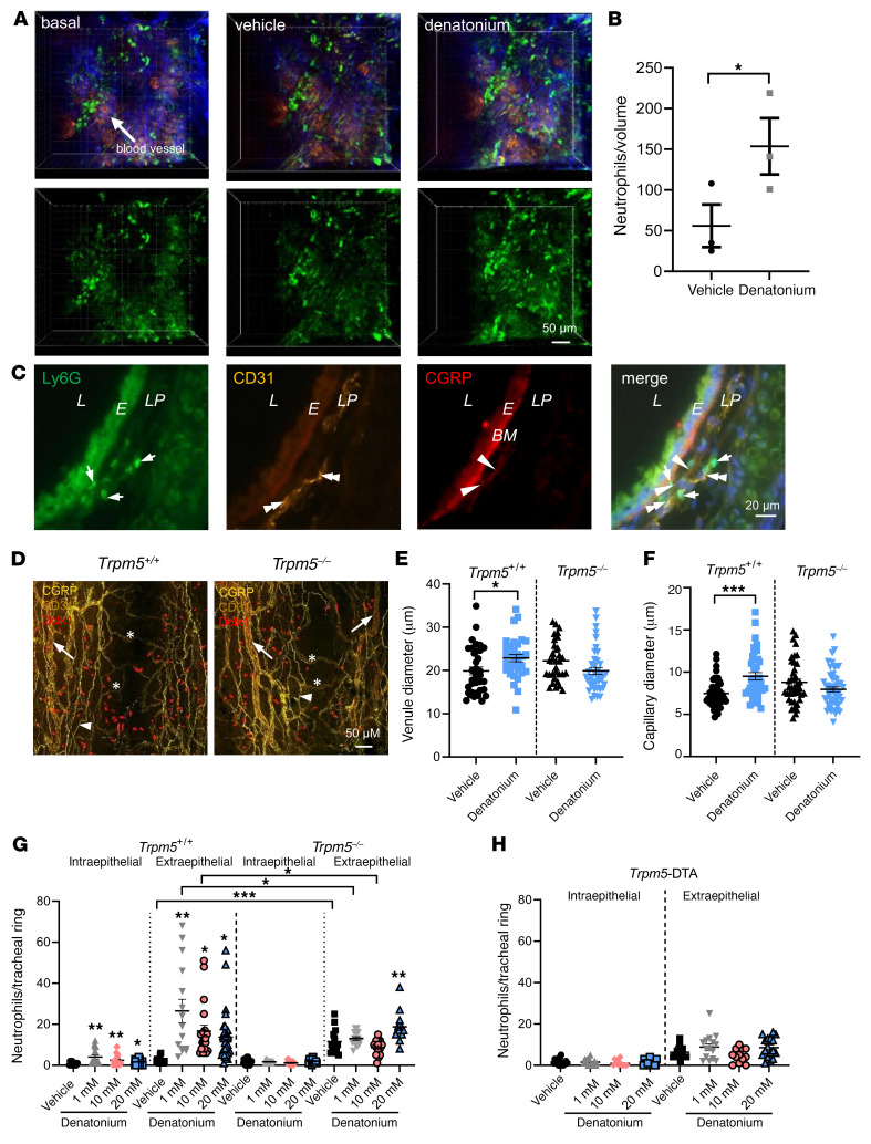
Figure 3. Denatonium evokes neutrophil recruitment and blood vessel dilation in the trachea.
(A and B) In vivo 2-photon microscopy of neutrophils and blood vessels in the trachea of Ly6G-GFP mice. (A) Denatonium-increased neutrophil (green) extravasation from blood vessels (red) (blue: second harmonic generation signal, collagen fibers) in WT mice compared to basal and vehicle-treated (HEPES-treated) controls. (B) Evaluation of the results in A (n = 3 mice). Volume: 300 × 300 × 60 μm. Denatonium: 20 mM. (C) Tracheae stained for Ly6G, CD31, and CGRP showed neutrophil recruitment (Ly6G, green) in proximity to blood vessels (CD31, yellow) and CGRP+ nerve endings (red) at the same site (merge). Evans blue bound to the basal membrane (BM, bright red). L, lumen; E, epithelium; LP, lamina propria. Merge: nuclei stained for DAPI (blue). (D) Whole-mount staining of tracheae from Trpm5+/+ and Trpm5–/– mice. BC: Dclk1 (red); blood vessels: CD31 (yellow), venules = arrows, capillaries = stars; nerves: CGRP (yellow, arrowheads). Scale bars: 50 μm (A and D) and 20 μm (C). (E and F) Quantification of the diameter of venules (E) and capillaries (F) (n = 32–53 vessels from 4 mice). (G and H) Analysis of neutrophils per tracheal ring in WT (Trpm5+/+) mice, Trpm5–/– mice, and BC-deficient Trpm5-DTA mice (n = 12–25 rings from 4–5 mice). In B and E–H, data are shown as single values and mean ± SEM. *P < 0.05; **P < 0.01; ***P < 0.001 by 2-tailed, unpaired Student’s t test (B) or 1-way ANOVA followed by Bonferroni’s multiple-comparison correction (E–H).

