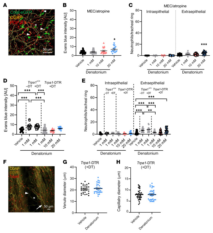
Figure 4. The denatonium-induced neurogenic inflammation is mediated via cholinergic signaling and sensory nerve activation.
(A) Nerve fibers (PGP9.5+: red) containing the neuropeptide CGRP (yellow) in Chat-eGFP mice (ChAT+ cells: green, stars). Arrowheads: neuroendocrine cells labelled for PGP9.5 and/or CGRP. Scale bar: 50 μm. (B) Quantification of Evans blue (EB) extravasation in response to 1, 10, or 20 mM denatonium in WT mice treated with the AChR antagonists mecamylamine (MEC) and atropine. (C) Analysis of neutrophil numbers per tracheal ring in WT mice treated with MEC and atropine. (D) Quantification of EB extravasation in response to 1, 10, or 20 mM denatonium in Trpa1-DTR mice treated with diphtheria toxin (DT) and in response to 1 mM denatonium in naive WT or DT-treated WT (Trpa1+/+) mice. (E) Analysis of neutrophil number per tracheal ring in response to 1, 10, or 20 mM denatonium in Trpa1-DTR mice treated with DT. Naive or DT-treated WT (Trpa1+/+) mice stimulated with 1 mM denatonium served as controls. (F) Staining of venules (arrows) and capillaries (stars) in tracheae from DT-treated Trpa1-DTR mice. BC: Trpm5 (red), blood vessels: CD31 (yellow), nerves: CGRP (yellow, arrowheads). Scale bar: 50 μm. (G and H) Quantification of the venule (G) and capillary diameter (H). n = 29–41 vessels from 4 mice. In B–E, G, and H, data are shown as single values and mean ± SEM (n = 14–24 rings from 3–4 mice). *P < 0.05; **P < 0.01; ***P < 0.001 by 1-way ANOVA followed by Bonferroni’s multiple-comparison correction (B–E) or 2-tailed, unpaired Student’s t test (G and H).

