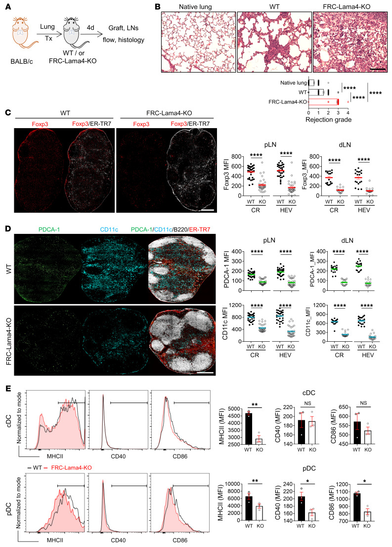
Figure 9. FRC-Lama4 regulates alloreactivity in lung transplants.
(A) Schematic of lung transplantation. BALB/c donors, C57BL/6 WT, or FRC-Lama4–KO recipients. Grafts and LNs harvested 4 days after transplantation. (B) H&E staining of native lung from BALB/c mice, lung grafts in WT and FRC-Lama4–KO recipients. Scale bar: 100 μm. Evaluation of rejection grade. (C) Whole-mount scanning of recipient pLN cryosections stained for Foxp3 and ER-TR7. Original magnification, ×20. Scale bar: 500 μm. Quantification of Foxp3 intensity in CR and around HEVs in recipient pLNs and lung dLNs. (D) Whole-mount scanning of recipient pLN cryosections stained for PDCA-1, CD11c, ER-TR7, and B220. Original magnification, ×20. Scale bar: 500 μm. Quantification of PDCA-1 and CD11c intensity in CR and around HEVs in recipient pLNs and lung dLNs. (E) Gating and summary of MHCII, CD40, and CD86 on cDCs and pDCs in WT and FRC-Lama4–KO recipient pLNs. (E) Representative of 2 independent experiments with 3 mice/group, 3 LNs/mouse, 3 sections/LN, 3 to 5 fields/section. Student’s unpaired, 2-tailed t test for 2-group comparisons. Data are represented as mean ± SEM. *P < 0.05; **P < 0.01; ****P < 0.0001. P < 0.05 was considered significant.

