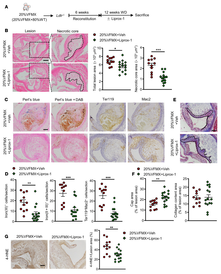
Figure 7. Liprox-1 alleviated accelerated atherosclerosis progression in Jak2VFMx-Cre clonal hematopoiesis.
20% of Jak2VFMX-Cre (VFMX) mice were fed with Western diet together with Liprox-1 (10mg/kg, 3 times per week) or vehicle injection for 12 weeks. (A) Experiment timeline. (B) H&E-stained images of aortic root sections. Necrotic core regions indicated by broken lines, and quantification of total lesion area and necrotic core area are shown. Scale bar: 200 μm. (C) Iron (Perl’s blue) and redox-active iron deposition (Perl’s blue + DAB); IHC staining of RBCs (anti-Ter119) and macrophages (anti-Mac2) in aortic roots. (D) Bar graph shows quantification of iron (II + III)–positive and erythrophagocytosis (Ter119+Mac2+) cell counts per section. Scale bar: 100 μm. (E) Aortic root sections were stained with Masson’s trichrome staining for fibrous cap (red, outlined by broken lines) and collagen (blue) content area, and then (F) quantified as the ratio of total lesion area. Scale bar: 100 μm. (G) Lipid peroxidation product 4-HNE staining, quantified as the percentage of total lesion area. Scale bar: 100 μm. Unpaired 2-tailed t test. *P < 0.05, **P < 0.01, ***P < 0.001.

