Fig. 4.

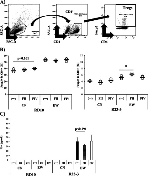
The hot water extract from G. hansenii GK-1 (FII) induced Foxp3+T cells in mesenteric lymph node (MLN) cells of R23-3 mice with severe inflammation caused by EW-feeding.
(A) Representative plots of a scheme examining the Foxp3+T cells. (B) Rates of Foxp3+T cells in CD4+T cells from MLN cells of RD10 (left) and R23-3 (right) mice fed with the EW-diet for 7 days (severe inflammation phase). MLN cells were incubated under Foxp3+T cell induction conditions stimulated with OVA (1 mg/mL), rIL-2, TGF-β, and retinoic acid, in the presence or absence of FII or FIV. The collected cells were analyzed by flow cytometry. (C) IL-4 production in the culture supernatant was analyzed by ELISA. 〇 indicates the value of each well, and the horizontal line indicates the mean of each group. The cells of 3 mice were pooled, cultured and analyzed for 3 wells for each condition. Significance was determined by Student’s t-test (*p<0.05, #p<0.1). The data are representative of two independent experiments.
