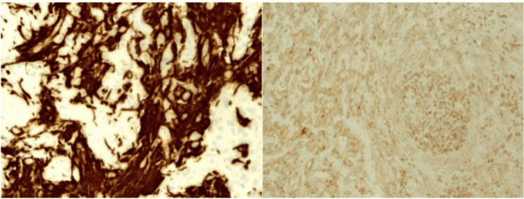
Figure 5. Left: Immunohistochemistry of glial fibrillary acidic protein (GFAP), located in the intermediate filaments of astrocytes. Right: Nuclear expression of protein gene product 9.5 (PGP 9.5), a member of the ubiquitin hydrolase family of proteins that is restricted to neural and neuroendocrine cells.
Tumor cells were negative for synaptophysin, Melan A, human melanoma black-45 (HMB45), and cytokeratin CAM 5.2. Cell proliferation index (Ki67) was 60% and phosphohistone H3 (pHH3) enhanced mitotic figures (up to 10 mitotic figures in 10 high-power fields). A second tissue sample sent as a biopsy of the left sacroiliac lesion showed metastatic involvement by a tumor with epithelioid and small cell characteristics similar to the images described, with positive tumor cells for protein gene product 9.5 (PGP 9.5) and negative for glial fibrillary acidic protein (GFAP), S100, SRY-box transcription factor 10 (SOX10), melanocyte inducing transcription factor (MITF). These markers confirm the cell lineage of the neoplasm [9].

