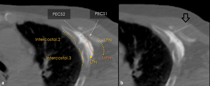
Fig. 1.

CT-guided PECS-2 nerve block. Images ( a , with annotations; b , without annotations) demonstrate needle placement (arrow, b ) and course of the relevant nerves in this axial CT image of the chest prior to ablation of chest wall tumor. LPN, lateral pectoral nerve; LTN, long thoracic nerve; MPN, medial pectoral nerve.
