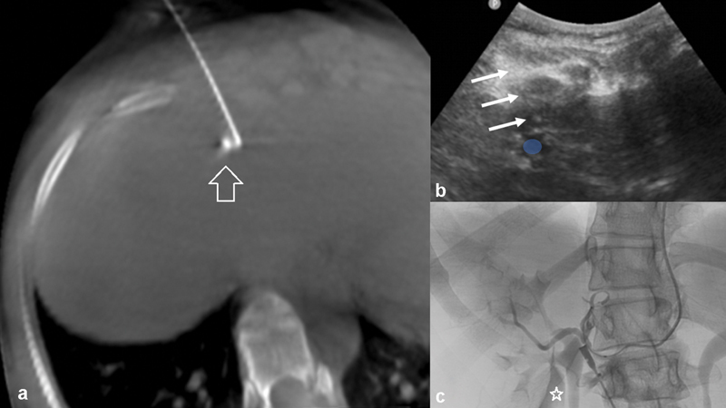
Fig. 5.

Ultrasound-guided and cone-beam CT-confirmed hepatic hilar block performed prior to left hepatic arterial bland embolization. Ultrasound guidance was used to target the left periportal space ( b , blue circle denotes left portal vein, arrows denote needle trajectory) and a small test injection was checked on plain X-ray to not wash into a vascular structure (also confirmed on cone beam CT arrow, a ). At this point, the nerve block is delivered. ( c ) Postembolization angiogram, with residual nerve block agent noted lateral to the renal collecting system (star).
