
Fig. 6.
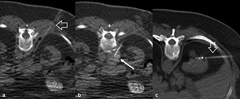
CT-guided aorticorenal plexus block prior to microwave ablation of kidney tumor. ( a ) Needle (arrow) trajectory. ( b ) The early injectate is seen spreading along the aorta and posterior to the renal artery (arrow) at the time point shown in this image. ( c ) Microwave antenna placement (arrow).