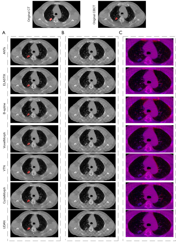
Figure 4.
The comparison of registration results between our method and the other mainstream methods. (A) The registered images, where the red area is the CBCT tumor segmentation label deformed by the deformation field; (B) the checkerboard display of registered images; (C) the red and blue channel overlay of the registered images, zoomed in on the lungs and parts of the surrounding area. CT, computed tomography; CBCT, cone-beam computed tomography.

