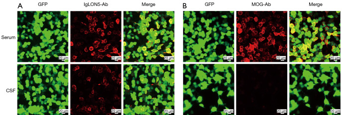
Figure 1.
Anti-IgLON5 and anti-MOG antibodies as shown in immunofluorescence tests using fixed cell-based assay. (A) Anti-IgLON5 antibodies 1:30 in the serum; Anti-IgLON5 antibodies 1:10 in the CSF. (B) Anti-MOG antibodies 1:32 in the serum. Scale bar =20 µm. GFP, green fluorescent protein; MOG, myelin oligodendrocyte glycoprotein; CSF, cerebrospinal fluid.

