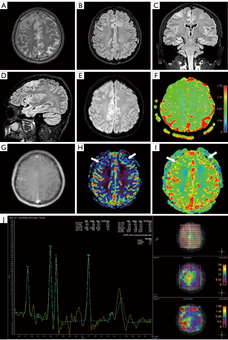
Figure 2.
MRI findings of the lesion. Brain MRI demonstrated cortex swelling and a hyperintense lesion on axial T2WI (A) and FLAIR (B-D) on the bilateral frontal and cingulate gyrus region. Mild hyperintensity on the axial DWI (E) and ADC (F) value decreased, bilateral frontal pia meninges showed contrast enhancement (G). CBF (H) and CBV (I) showed decreased blood flow in the bilateral frontal and cingulate gyrus (white arrows). MRS (J) demonstrated a lower peak of the NAA signal and higher peaks of the Cho and Cr signal (Cho/Cr: 1.38; Cho/NAA: 1.17; NAA/Cr: 1.17). MRI, magnetic resonance imaging; T2WI, T2-weigted image; FLAIR, fluid attenuated inversion recovery; DWI, diffusion-weighted imaging; ADC, apparent diffusion coefficient; CBF, cerebral blood flow; CBV, cerebral blood volume; MRS, magnetic resonance spectroscopy; NAA, N-acetyl aspartate; Cho, choline-containing compounds; Cr, creatine derivatives.

