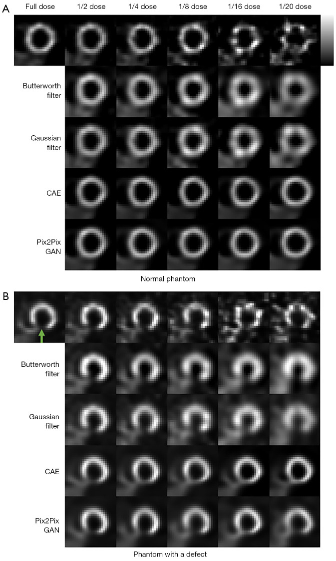
Figure 7.
Simulated MP SPECT images with various dose levels of (A) a sample normal male phantom and (B) a sample abnormal female phantom processed under different denoising methods. The green arrow indicates the defect location. CAE, convolutional auto encoder; GAN, generative adversarial network; MP, myocardial perfusion.

