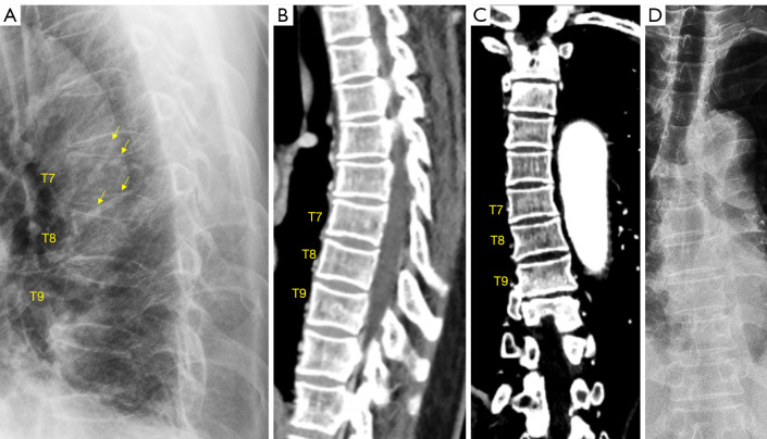
Figure 10.
Chest imaging of an elderly woman. Lateral radiograph (A) mimics of both upper and lower endplate depressions of vertebrae T7, T8, and T9. Careful observation shows the endplate rings are projected as apparent ovals (arrows), suggesting “rotation” of vertebrae relative to expected position (or relative to X-ray beam). Reconstructed sagittal CT image (B), coronal CT image (C), and frontal view radiograph (D) show slight scoliosis of T6-T9 vertebrae (toward right side). There is no apparent VD for T7-T9 vertebrae on images of (B-D). Note the anterior vertebral heights of T7–T9 vertebrae appear maintained on (A). Reproduced with permission from (17). VD, vertebral deformity.

