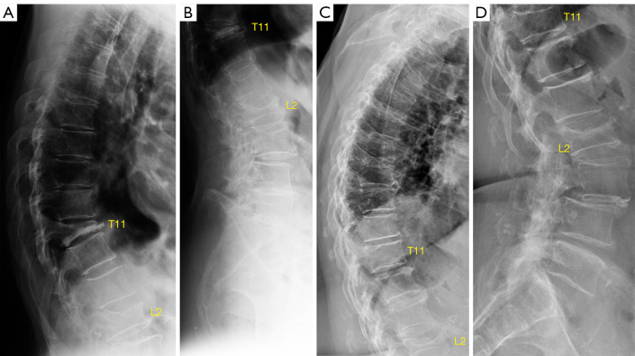
Figure 11.
X-ray projection has important implications in the appearance of vertebrae. (A) and (B) are from one elderly woman, and (C) and (D) are from another elderly woman. (A,B) show vertebra T11 is collapsed. Note the extent of L2 vertebral height loss appear to be more severe in (B) than in (A). It is likely that L2 is more ‘off-center’ to the X-ray beam focus in (B) than that in (A). T11 upper endplate shows depression mimics on (D), while on (C) T11 upper endplate appears to be normal. Note the extent of L2 vertebral height loss appear to be more severe in (C) than in (D). It is likely that L2 is more ‘off-center’ to the X-ray beam focus in (C) than in (D).

