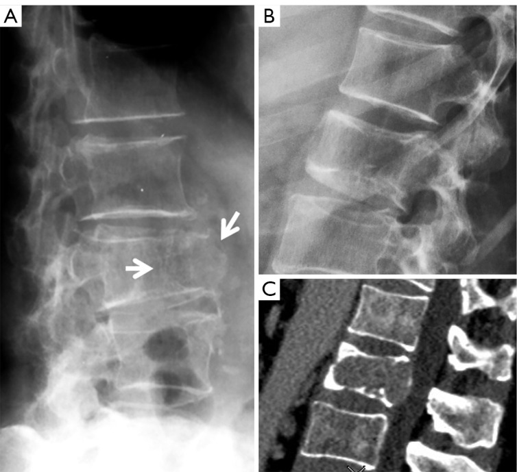
Figure 13.

Two cases of oncological vertebral deformities which can be easily differentiated from OVF. Radiograph (A) of a 68-year-old woman shows osteolytic pathological fracture of L3 level due to malignant tumor (arrows). The lesion shows loss of integrity of bone cortex, trabecular network and connectivity at the anterior vertebral body with destructive and expansive features. The anterior upper corner of lower vertebra (L4) is also destroyed. Radiograph (B) and CT (C): a case of L2 vertebral metastasis of lung carcinoma. L2 shows height loss and both upper and lower endplate depression. Lower endplate demonstrates irregular shapes which differ from osteoporotic depression. Reused with permission from (15). OVF, osteoporotic vertebral fracture.
