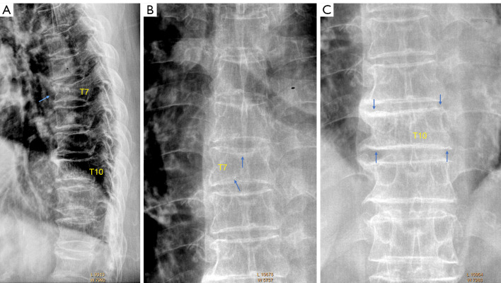
Figure 2.
Typical bi-concaved OVF. A typical OVF is shown on (A). In addition to its typical location in the mid-thoracic location, both upper and lower endplates are depressed, and anterior cortex buckling is also noted (arrow). T7 upper endplate and lower depressions (arrows)are also seen on frontal radiograph (B). Vertebral T10 is what we called ‘short vertebra’, with the anterior height and posterior height reduced to a similar extent. (C) Confirms OVF of vertebral T10 (arrows). OVF, osteoporotic vertebral fracture.

