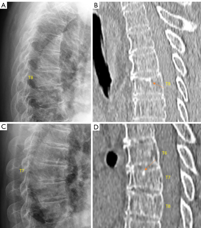
Figure 4.
Chest imaging of two elderly women (A, B for one patient and C, D for another patient). Radiograph (A) shows T8 vertebra with minimal wedging without definite upper endplate depression. CT (B) shows T8 height loss and apparent upper endplate depression (arrow). Radiograph (C) shows T7 vertebra with mild wedging without definite upper endplate depression. CT (D) shows T7 height loss and apparent upper endplate depression (arrow). (A) and (B) are reproduced with permission from (17). CT, computed tomography.

