Figure 7.
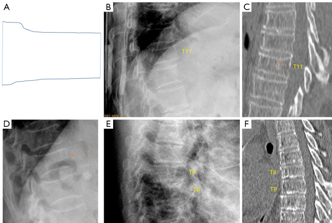
Short vertebra. (A) A line drawing of a short vertebra. We define short vertebrae as those with middle height and anterior height reduced to the similar extent. (B,C) Chest imaging of an elderly woman shows T11 short vertebra. (B) Radiographic appearance. Endplate depression (arrow) is present on CT (C), suggesting this deformity is likely to be osteoporotic. (D) Radiograph of an elderly woman shows T11 short vertebra (arrow), we consider it be osteoporotic (no confirmation was obtained). (E,F) Chest imaging of an elderly woman shows T8 and T9 appears as short vertebral height, while no endplate depression is noted. No endplate depression is noted on CT (F) for T8 and T9. Endplate sclerosis is considered for vertebrae T8, T9, T10 on CT (F). T8 and T9 short vertebrae are likely due to ‘osteoarthritic’ changes. (B,C,E,F) are reproduced with permission from (17). CT, computed tomography.

