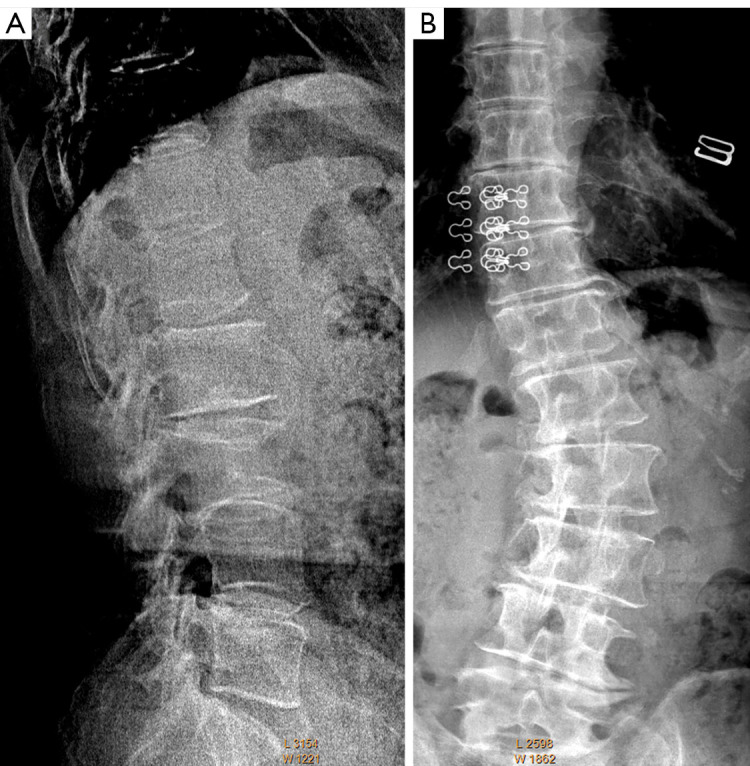
Figure 9.

A case of scoliosis in an elderly woman. (A) Lateral radiograph shows L3 OVF mimics. (B) Frontal radiograph shows lumbar spine scoliosis, however lumbar vertebrae appear to be of normal shape. Note the anterior height of L3 appears maintained. OVF, osteoporotic vertebral fracture.
