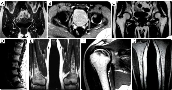
Figure 2.
MRI images diversity of infiltration patterns and complications in bone marrow. [The images are provided by Dr. Mercedes Roca and are part of her publications “Magnetic Resonance Imaging of Bone Involvement in Gaucher Disease” ISBN 978-84-945315-7-6 (18) & “Resonancia Magnética en Enfermedades Hematológicas” ISBN 84-7885-275-1) (3). The publication of these books was her own initiative and that the images are of their property and they were not ceded to the publishers who published these books, with whom she had not signed any copyright agreement because the books were not copies for sale.] (A) SE T1 WI Coronal pelvis: Homogeneous pattern. No sign of fatty marrow. (B) SE T2 WI axial hips. Initial ischemic focus on the left femoral head. (C) SE T1WI Coronal hips: small foci of hematopoietic marrow around the lesser trochanter, in pelvis with abundant fatty marrow. (D) SE T1 WI Sagittal lumbar. Low signal infiltrative foci. Melanoma metastasis. (E) SE T1 WI Coronal distal femur. Medullary infiltration as mottled foci with preserved epiphyses. (F) SE T1 WI Coronal T1 shoulder: Bone infarction. Intramedullary ischemic area. (G) SE T1 WI Coronal tibias. Non-homogeneous reticular pattern. MRI, magnetic resonance imaging; SE, spin echo; T1 WI, T1 weighted imaging; T2WI, T2 weighted imaging.

