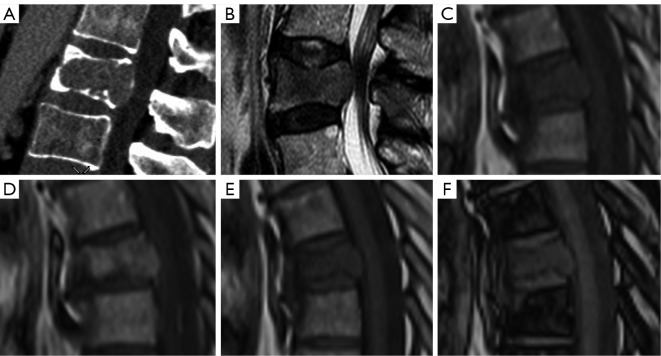
Figure 11.
Pathologic fracture in several cases of metastatic lung cancer. Sagittal CT (A) and sagittal T2 weighted images (B) showing convex posterior border. Sagittal T1 weighted image without (C) and with (D) contrast in pathologic fracture. In phase (E) and out of phase (F) showing lack of fat in this metastatic vertebra, with a signal intensity out phase/phase ratio of 0.95. CT, computed tomography.

